One inevitable outcome of aging is that the body starts to deteriorate. Everyone ages differently, but everyone ages. For me, the knees are the first casualty of time.
My right knee has been bothering me for many years now, but recently the pain has become more frequent. After putting it off forever, I finally decided to get it checked out by an orthopedic doctor.
The waiting room was filled with people, including those with arms in casts, legs in braces and one lady in a wheelchair. My consultation took only 15 mins, with the doctor taking note of my medical history followed by a physical exam of the knee.
He ordered X-Rays and MRI scans, which I completed later that afternoon in the same hospital. Very efficient, and the review appointment was confirmed for a week later.
But first, here’s the basic anatomy of your typical knee.

Looking deeper, we find many ligaments that connect the various knee bones and cartilage that provides lubrication between them. Specifically, I’ve highlighted (in red) the Articular cartilage, Anterior cruciate ligament (ACL) and Quadriceps tendon.

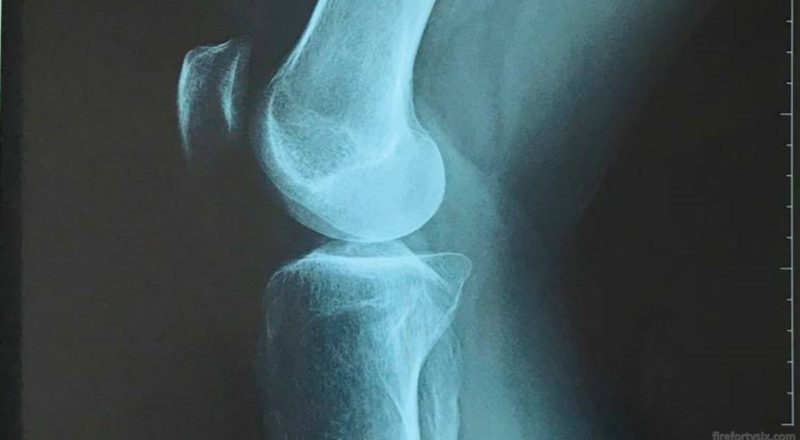
During the review session, the doctor started with X-Ray films which showed a “slight reduction in the medial tibiofemoral joint space”. Which in English means that the space in-between the tibia and femur, on the inside part of the knee, is slightly less than expected.


This observation is a common result of osteoarthritis, where the Articular cartilage has been worn down, causing pain in the knee joint. Since X-Rays only capture bone and not cartilage, this was not immediately visible but it was confirmed with the MRI scans.

The two additional observations from the MRI scans were a “strain and partial tear of the right ACL”, which means exactly what it sounds like; and “right quadriceps tendinosis”, which is a degeneration of the collagen in the quadriceps tendon.

Nothing particularly serious, and no invasive surgical procedures required at this time. However, the doctor advised me to avoid strenuous sports that involve knee twisting (like badminton) and bending (like cycling), and to minimise climbing stairs.
Surprisingly, he was fine with running, but only after the right quadriceps was sufficiently strengthened with exercises like leg raises. He wrote up a referral note for follow-up physiotherapy, and wished me well.
I asked him about the effectiveness of popular dietary supplements like glucosamine, chondroitin, MSM and UC-II, and his view was that they didn’t provide any clinical benefit. They don’t have adverse effects and can sometimes provide anti-inflammatory, so it was down to personal choice whether to take them.
I’ve been wearing knee support braces for quite some time, and asked if I should continue. “Sure, why not”, was his reply. Here’s my current collection, and as you can see, I’m partial to the widely-available LP brand.


It was reassuring to know that my condition wasn’t too serious and was fairly common for men my age. I will have to live with the chronic knee pain though, but in the grand scheme of things, it’s not that bad and life goes on.